 ×
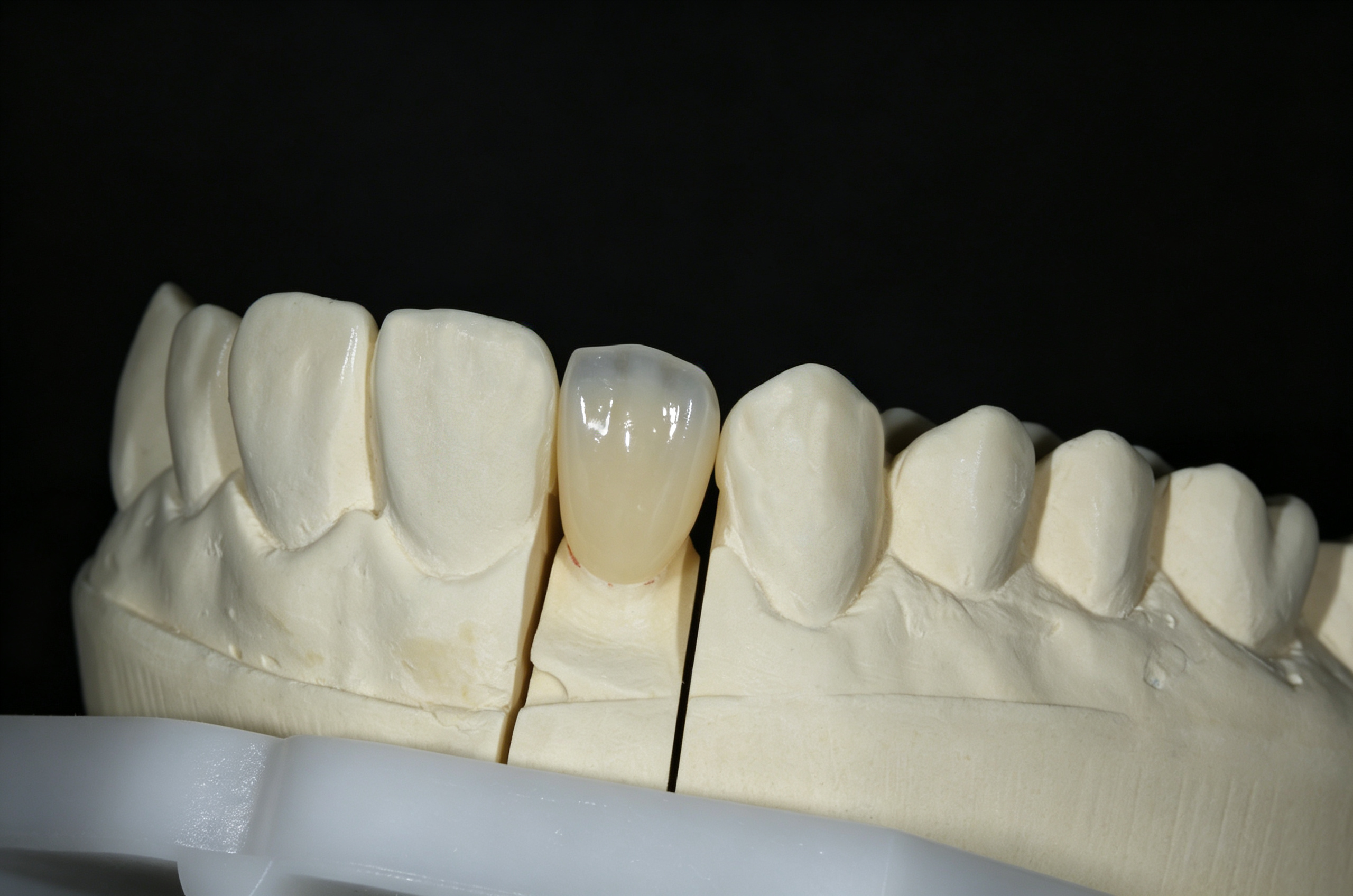
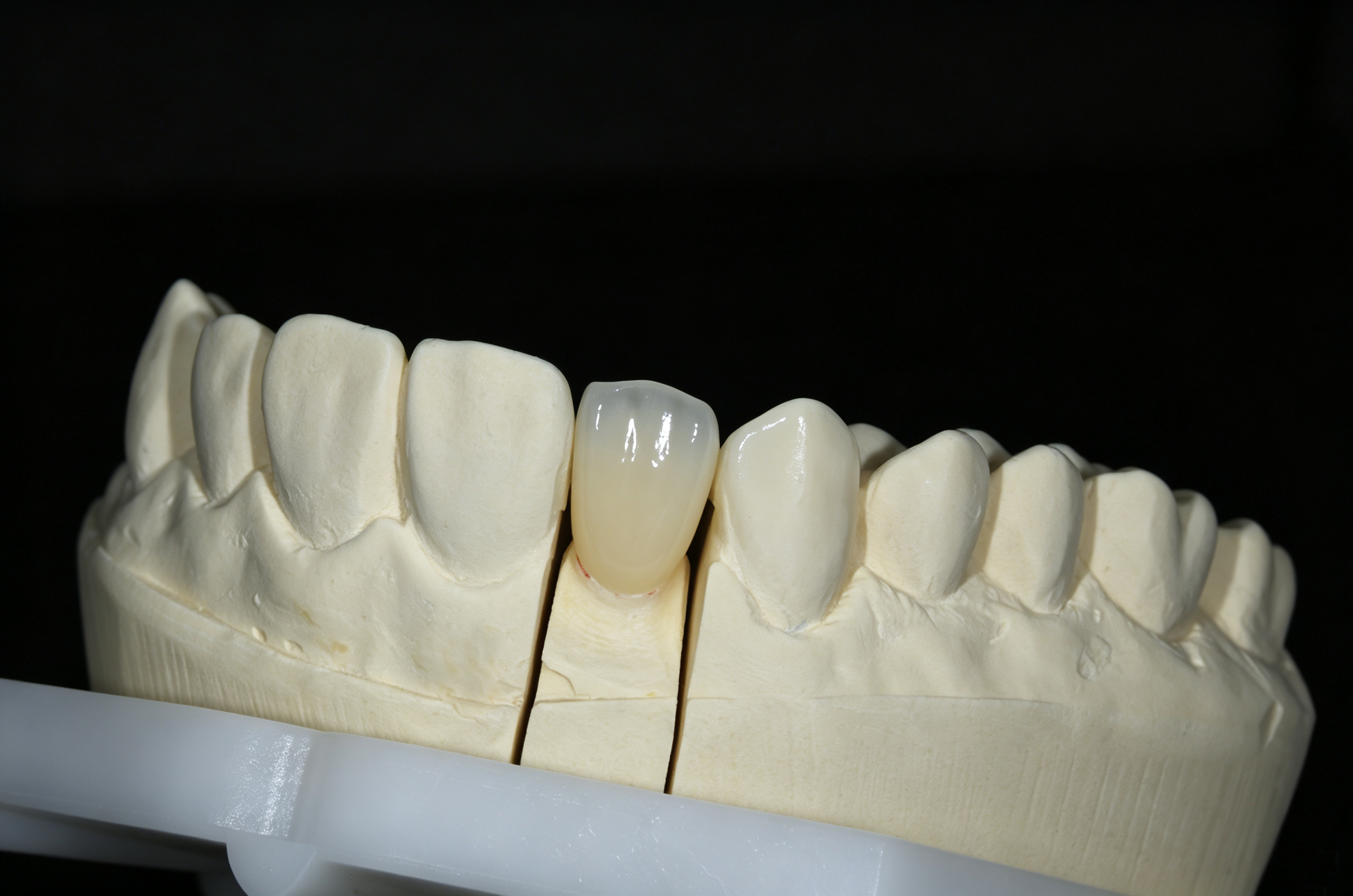
This anterior single crown was made with ICERA 4D zirconia and photographed on a sectioned stone model. Its warm cervical and body tones transition to a cooler incisal appearance, while a tapered outline and rounded contours define its shape. Close-u...
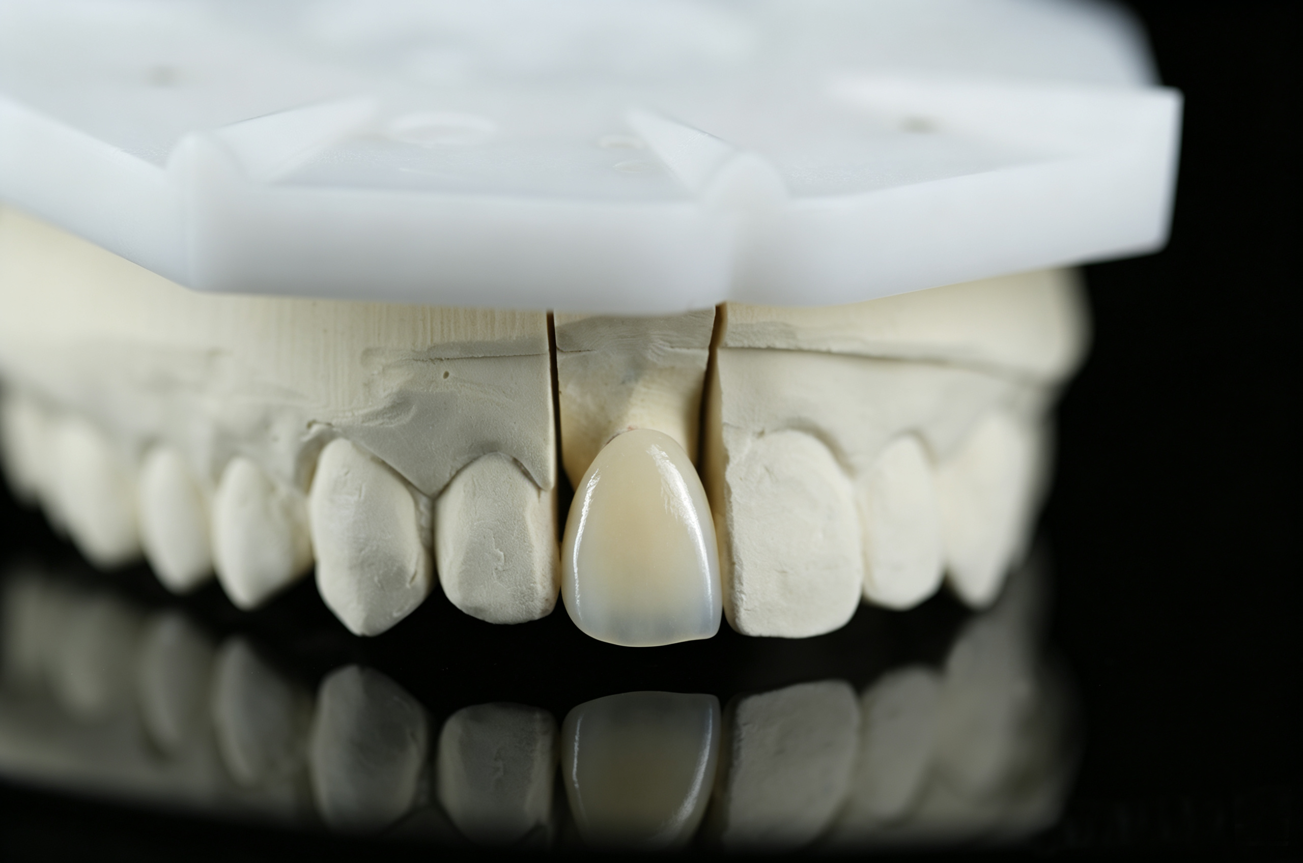
This anterior single crown was made with ICERA 4D zirconia and photographed on a sectioned stone model. Its warm cervical and body tones transition to a cooler incisal appearance, while a tapered outline and rounded contours define its shape. Close-up views highlight the glossy facial surface and the crown’s position among the adjacent model teeth. The case offers a visual reference for laboratories and partners exploring ICERA 4D zirconia for anterior restorations.
An anterior restoration offers a close view of how shape, color, and surface appearance work together. In this case, a single crown made with ICERA 4D zirconia is presented on a sectioned stone model. Three laboratory photographs show the restoration from slightly different facial perspectives, with the surrounding model providing a reference for its position and outline.
The crown has a warm cervical and body color, a cooler incisal zone, and a glossy facial surface. Its outline broadens toward the incisal portion and narrows toward the cervical area. These visible details form the focus of this case: a finished zirconia crown viewed within the anterior segment of a laboratory model.
A single crown within the anterior segment
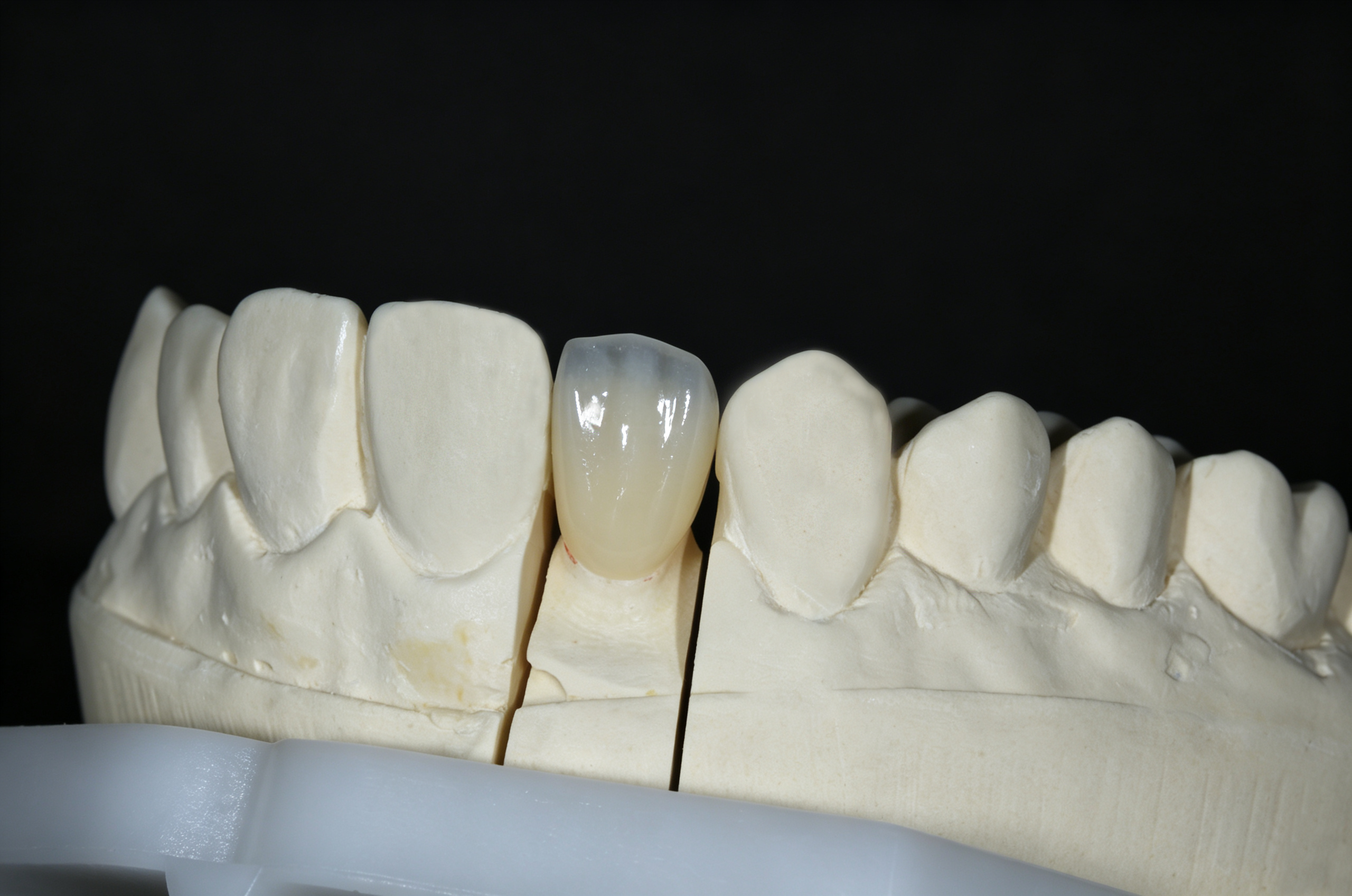
The finished crown sits between a broad incisor form and a more pointed canine form on the model. Its narrower outline gives it a distinct identity within that sequence. The incisal edge is gently curved, with rounded transitions into the proximal outlines. Toward the cervical area, both sides converge into a slimmer neck.
This shape is easiest to read when the crown is viewed together with its immediate neighbors. The adjacent stone teeth establish a sequence of different widths and silhouettes, while the finished crown remains clearly identifiable through its color and reflective surface. The photographs therefore show both the individual restoration and its relationship to the surrounding modeled anatomy.
The facial surface appears softly convex. Its central area is relatively broad, with rounded transitions toward the sides. The overall impression is of a smooth contour with restrained anatomical detailing.
From cervical warmth to a cooler incisal appearance
The most noticeable color feature is the difference between the lower and upper portions of the crown. The cervical region has a warm cream tone, which continues through the body. Moving toward the incisal edge, the appearance becomes cooler and more gray-blue in the photographed lighting.
This cooler zone is visible across all three images. It gives the incisal portion a different visual character from the warmer body beneath it. Against the dark background, the upper outline and the change in appearance are particularly easy to follow. The crown reads as several connected regions rather than one uniform color.
The transition is broad, with the strongest contrast concentrated near the incisal portion. The middle region links the cooler edge to the warmer cervical area. For readers examining the photographs, the useful reference is this distribution of color across the crown. The surrounding stone provides anatomical context; it does not provide a reference for matching the shade of natural teeth.
Surface detail through reflected light
The crown's facial surface carries several bright vertical reflections. Their positions change between photographs, allowing different parts of the contour to become visible. The glossy finish contrasts with the matte, textured surface of the stone model and draws attention to the restoration at the center of the image.
In areas between the strongest reflections, the facial surface looks relatively smooth. The visible character is defined mainly by the broad contour and color transition, with little pronounced surface texture apparent in these views. The reflections also cover portions of the surface, so the photographs offer a clearer record of the overall outline than of very fine texture.
The images document the surface as photographed. The specific finishing technique has not been recorded for this case, so no particular staining, glazing, polishing, or veneering sequence is attributed to the crown.
Reading the crown on a sectioned model
The sectioned model makes the individual crown and its supporting die easy to distinguish. Separation lines on either side of the die remain visible beneath the restoration. This laboratory setting gives the photographs a clear context and keeps attention on the crown's external form.
The closer facial view provides the clearest overview of the incisal outline, body color, and cervical taper. The slightly more oblique view adds another perspective on the rounded facial contour and the way the surface catches light. Together, these views provide a useful visual record without requiring three nearly identical images in the main gallery.
These are laboratory photographs of a crown on a model. They document its visible appearance at that stage; clinical fit, bite relationships, and the final appearance in the mouth are outside the scope of this image set.
ICERA 4D zirconia in a finished restoration
The material used for this crown has been confirmed as ICERA 4D zirconia. This gives the case a direct connection to the material behind the finished restoration. The photographs let readers examine an actual crown made from that material, alongside the product information they may review separately.
The specific shade and fabrication method have not been documented here. The warmer cervical region and cooler incisal appearance are therefore described as features of this finished crown. They are not used to identify a shade code or reconstruct the technician's process.
For laboratories and distribution partners, that distinction supports a more useful material discussion. The case supplies a visual reference for an anterior application, while questions about product selection, processing requirements, and individual case planning can be addressed with the relevant technical information.
A reference for the next conversation
When discussing the appearance shown here, three details provide a practical starting point: the width and taper of the crown, the extent of the cooler incisal zone, and the degree of gloss visible on the facial surface. Identifying these details helps make a reference photograph more specific than a request for a generally natural-looking crown.
This case brings those features together in one anterior restoration made with ICERA 4D zirconia. The photographs give laboratories and potential partners a close view of the finished crown and a concrete basis for discussing the material.
To learn more about ICERA 4D zirconia, contact our team with your intended restoration type and material requirements. Reference this case if its contour or color distribution is relevant to your inquiry.


 EN